5.16.2011

ELISA (tbe)

Overview
A pathogen entering the body stimulates the immune response. The body responds to the invading organisms, or antigens, by producing antibodies. These antibodies act as flags, binding to the antigens and signaling other cells to come and destroy them. Antibodies also remain in the bloodstream after the pathogen is destroyed, ready to react if the pathogen returns a second time. Because each antibody is specific to a certain antigen, they can be used to detect the presence of a disease in an individual.

Procedure
The Enzyme-Linked Immunosorbent Assay, or ELISA, can be used to test for antibodies. It has multiple uses, such as testing for diseases such as HIV and SARS, drug use, and pregnancy. In ELISA, first a test sample and then a primary antibody solution are added to a microplate strip. If antigens are present in the test sample, they will have bound to the wells on the microplate strip, and antibodies will bind to those antigens. If not, they will be removed when the well is rinsed out. A second antibody is then added to the microplate strip, and the unbound antibodies are again rinsed out. The second group of antibodies are labeled with an enzyme which, when exposed to a chromogenic enzyme substrate, will change color.
                                                        The color-changing enzyme shows up
                                                         as the second band on a pregnancy test.

4.11.2011

Sushi Lab

Background

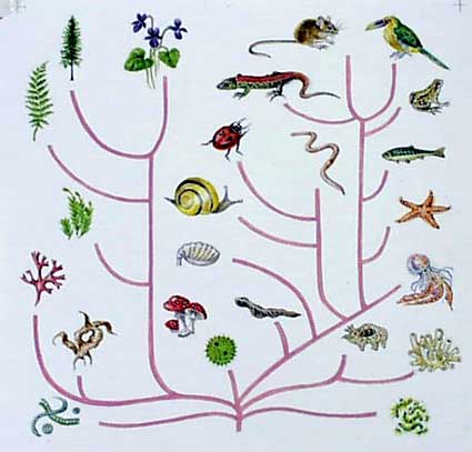
As far as we know, all life on Earth evolved from a single common ancestor. Therefore, all organisms are related to a certain extent. However, the closeness of relations between various species, and the point in their evolution when they split off from each other, is still unknown. In the past, scientists traced evolution by using anatomy to identify similar structures, and used this to build an evolutionary tree. However, this method has its shortcomings. For example, although chimpanzees look more similar to gorillas than they do to human beings, they are actually more closely related to us. With the use of Biotechnology, Proteomics- the study of proteins and their functions- allows researchers to trace how species have evolved in a much more precise and accurate manner.


Procedure
The fish muscle samples are being incubated for five minutes at room temperature in the sample buffer. The resulting mix (sans fish) is poured into a microtube and heated for five minutes at 95°C. This denatures the protein, unraveling it so it can be organized by gel electrophoresis. After GE, the samples will be arranged in bands according to size. The more similar bands are between samples, the more closely those two species are related.

3.28.2011

Mitochondrial DNA

Background
In addition to nuclear DNA, each cell contains mitochondrial DNA, suitably named for its location in the mitochondria. Mitochondrial DNA is used to create and store ATP, which is used to power the cell. It is theorized that mitochondria may have been bacteria adapted into the cell in a symbiotic relationship; the bacteria would receive nutrients from the cell in return for energy created from oxygen.
^ Mitochondria_______________________

Mitochondrial DNA is inherited only from the mother, because while the sperm and oocyte both contribute chromosomes, only the oocyte contributes cytoplasm and organelles. Mitochondria also have a high mutation rate- approximately 10 times as often as nuclear DNA. These two features make it possible to trace ancestry by comparing the mutations present in individuals to those of their mothers, grandmothers, great-grandmothers, etc., etc. Work in the 1980s by Alan Wilson and others at UC Berkeley created a family tree, hypothesizing that all humans alive today evolved from a single ancestor. This "mitochondrial Eve" probably existed in Africa about 200,000 years ago.
^ a family tree_________________________

Mitochondrial DNA is extremely valuable in science. Because it is more prolific than nuclear DNA, it can be used in cases where much time has passed and DNA has been severely degraded.
Procedure
Skin cells are obtained either by a saline mouthwash or root ends from scalp, eyebrow, or arm hairs. A region is amplified using PCR, which cycles the DNA through three stages: first, a 94°C incubation to denature the DNA, a 58°C incubation to bond the primers to their complementary sequences, and a 72°C incubation for DNA polymerase to create complementary strands. Because of the large quantity of mitochondrial DNA already present, it does not need to be cycled as long as nuclear DNA. Because of the similarity of mitochondrial DNA, the bands resulting from gel electrophoresis will appear to be the same. Samples may be submitted to the Sequencing Service  of the Dolan DNA Learning Center, which can generate sequences and post them on the internet.
Results
From our results, we saw that the band width and positions are nearly identical to the human eye. This is understandable, as all human beings on the planet today share 99.9% of their DNA. The similarity allows us to trace our evolution and understand how we compare to each other, as well as to other species.
The next step in this lab will be sending our samples in to be sequenced, so we can compare our DNA in greater detail.

3.15.2011

"Disease" Gene Testing

Background
DNA testing has multiple purposes, and is used in the study of evolution, as well as both forensic and paternal tests. One of its most valuable applications is checking for genetic diseases. Through DNA testing, one can test whether an individual is at risk for inheriting an illness such as cystic fibrosis or sickle cell anemia. Scientists take a sample of cells, amplify them using PCR, and analyze them through gel electrophoresis. In this lab, we will use the above steps to test for a gene (which is actually an intron. This is due to the ethical problems of testing for genetic diseases in class).
Procedure
Day 1: Collecting a Sample
A sample of skin cells is taken from the subject by chewing gently on the insides of the cheeks and then rinsing with a saline solution. The sample, once collected in a test tube, is spun in a centrifuge to clump all the cells together. The test tube is then immersed in two hot water baths, one at 56°C and one at 95°C. This is done to break the cell membrane and release the DNA. Once loose in the cytoplasm, however, DNAse would quickly destroy the sample DNA. This is why Instagene Matrix beads are added to the sample before it is immersed in the hot water baths. The test tube is then stored in a refrigerator until the next day.
Day 2: Polymerase Chain Reaction (PCR) Amplification

The sample of cells taken from an individual's cheek is much too small to be visible using gel electrophoresis (discussed during Day 3). PCR is therefore necessary in order to increase the size of the DNA sample before gel electrophoresis. In PCR, a primer is added to the solution. This primer acts like a heat-seeking missile, searching for the target gene sequence. The sample is then placed in a thermal cycler, which cycles through three temperatures- one to denature, or "unzip" the DNA, one to anneal to (find) their complementary sequences of DNA, and one to make new nucleotides attach to their complements on the unzipped strands. These three steps constitute one cycle; one PCR amplification consists of 40 of these cycles.
Day 3: Gel Electrophoresis
The bands of DNA are separated by size during gel electrophoresis, with the smaller bands being further towards the bottom (having progressed faster than the larger ones). There are three possible results.
If the gene being tested for is present in both chromosomes, a band of DNA strands 941 base pairs long will form. If it is present in neither, a band of DNA strands 641 base pairs long will form. If the gene is present in one chromosome but not the other, one band of each will form.
Results
There was a major source of human error for our result, which was that the gel was punctured by the pipette tip one two or three wells and somehow all of the samples bled through to some extent. All of our control samples were barely visible viewed in the correct light, as well as Lizzie's results (she was heterozygous, meaning she carried the 'disease' but did not herself show symptoms). Over half the class's results were homozygous negative, meaning they had the disease, so although our other test samples did not yield results, it would not be unreasonable to hypothesize that at least two of our tablemembers tested homozygous negative.

2.01.2011

Testing for Genetically Modified Organisms Through the Use of PCR

   Background
What are GMOs?
GMOs, or genetically modified organisms, are animals or plants that have had traits added or amplified for human benefit. Genetic modification can be as simple as selective breeding of a species in order to bring out a quality. However, in recent years it has become possible to insert a gene from one species directly into another species. The most common uses for GMOs are in the agriculture business. Crops are often modified to improve features such as frost, pest, or drought resistance, to give plants longer shelf life, or to improve appearance and taste.
How are They Made?
The GOI (gene of interest) is identified in one species and isolated. This gene is placed in a plasmid, which is inserted into an agrobacteria. The agrobacteria is then inserted into a plant cell. The cell sees the inserted gene as one of its own, and the plant will develop with the specified trait.
Identification
There are two methods used to identify GMOs: ELISA and PCR. ELISA (enzyme-linked immunosorbent assay) identifies genetically modified organisms by identifying specific proteins. The disadvantages of this method are that the produce being tested must be fresh, and each test must be specific to a certain crop. However, ELISA is inexpensive and relatively simple. PCR (polymerase chain reaction), on the other hand, identifies all sequences of DNA foreign to the plant. Because a relatively small number of DNA sequences are inserted into crops, it is easy to test for many at once (a PCR test can identify up to 85% of all GM crops).
Controversy
GMOs have both advantages and disadvantages. Positive effects include the reduction of pesticides and herbicides in agriculture, and the possibility of nutritional value and reduced stress on farmland. However, opponents of GMOs warn that "superweeds" and "superpests", no longer affected by resistant crops, could develop. Other fears include reduction of plant diversity and potential allergic reactions resulting from crossing species. Although there are requirements in Europe to label genetically modified foods, there are no such laws in the United States due to the influence of agribusiness lobbies.
Procedure
1. Put 1 gram of cornflour into a mortar. Add 10 ml of distilled water, and grind into a pipetable mixture. Repeat with the broccoli. This is done to break down the cell wall.
The cornflower supplied in this lab is a non-GMO control. It verifies that the test will not give results for non-GMO foods with GMO primers.
2. Pipet 50 μL of the cornflour mixture and the broccoli into their respective tubes (labeled "non-GMO" and "test"). Add 50 μL of InstaGene to each tube.
3. Place the tubes in a 95°C water bath for five minutes. This breaks the cell and nuclear membranes. Afterwards, centrifuge the tubes.

The InstaGene added in step two prevents DNAse in the cytoplasm from destroying cell DNA that is released once the nuclear membrane is broken.
4. Prepare six test tubes. The contents should be as follows:

  1. 20 μL non-GMO control (cornflour), 20 μL plant master mix
  2. 20 μL non-GMO control, 20 μL GMO master mix
  3. 20 μL test food (broccoli), 20 μL plant MM
  4. 20 μL test food, 20 μL GMO MM
  5. 20 μL GMO positive control, 20 μL plant MM
  6. 20 μL GMO positive control, 20 μL GMO MM
The master mixes are primers which will search for a specific sequence of DNA. Because tubes one, three, and five contain plant primers, they should not show up as bands after the PCR testing. Tube two contains GMO primers, but the food is guaranteed to be non-GMO. If a band shows up from the test, this indicates contamination or some other failure in the experiment. On the other hand, tube six must produce a band, because it contains the GMO-positive control with GMO primers. A lack of a band would also mean a failure in the experiment. Tube four is the only one with completely unpredictable results.
5. Place tubes in the PCR machine (thermal cycler). The PCR machine uses fluctuating temperatures to replicate DNA.

6. Load 20 μL of each sample into the gel electrophoresis apparatus and run the gel for 3 minutes at 200 V.

Results


Bands appeared in lanes 2, 4, and 6. This means that the test food, broccoli, is genetically modified. There is an extra band in lane 2, the cornflour, which is likely due to contamination during the procedure.

1.25.2011

pGLO Transformation

Background
Genetic transformation is a process which entails taking a gene from one organism, and inserting it into another so as to alter or add a trait. Uses of genetic transformation include modifying plants to resist frost or pests, strengthening agriculture; altering bacteria to help in environmental disasters by giving them the capability to digest oil; and gene therapy, in which sick cells are transformed with the use of healthy genes.
To move genes from one organism to the other, the gene must be isolated and added to a plasmid. Plasmids are a small circular pice of DNA in addition to the chromosome. They allow bacteria to share genes and help them adapt to their environment more rapidly. To force the bacteria to accept the DNA, the plasmids and bacteria are subjected to heat shock, or a rapid raising and lowering of temperature. This triggers the bacteria's' instincts to absorb new DNA, a survival skill.
This lab involves taking the Green Fluorescent Protein gene from jellyfish and inserting it into bacteria, which will cause them to become bioluminescent.